Quick Summary
Additional Context
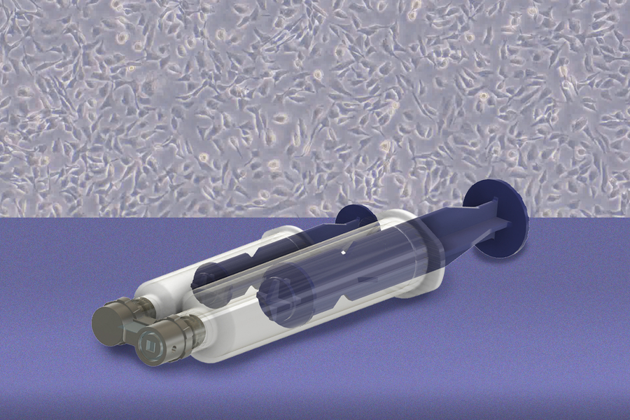
Researchers at the Massachusetts Institute of Technology (MIT) and Johns Hopkins University have developed a handheld tool built around a 3D printed microfluidic device. It collects living cells from selected spots on freshly excised tissue and leaves the rest of the tissue available for pathology. The team says the method could support earlier detection of ovarian cancer and many other cancer types, and could eventually be used to test how an individual patient’s cells respond to treatment. The findings appear in the journal Device.
Ovarian cancer is among the deadliest forms of the disease, in large part because it is often diagnosed late. Many cases begin in tissue that is difficult to sample at an early stage.
Where the Disease Starts and How Tissue Is Processed
Five-year survival f